The Ultrafast and Nonlinear Optics Lab at Penn State![]()
The Ultrafast and Nonlinear Optics Lab at Penn State![]()
Chromatic Imaging
Chromatic Confocal Imaging
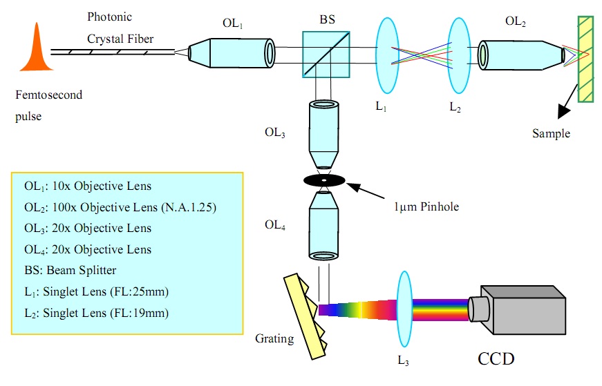
We have demonstrated a novel chromatic confocal microscope system using supercontinuum white light generated from a photonic crystal fiber. The chromatic aberration of a pair of singlet lenses is employed to focus the different spectral components of the supercontinuum at different depth levels. An effective depth scanning range of 7 µm is demonstrated. The corresponding depth resolution is measured to be less than 1 µm (FWHM).
Figures adopted from Shi et
al. (2004) [1].
Figure
top right: Confocal microscope system.
Figure
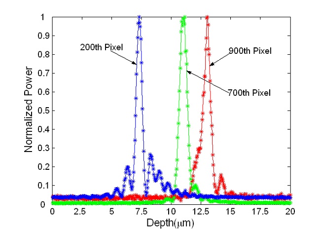
side right: Typical depth response curves. These curves show the dependence of the
reflected light intensity on depth position at a given wavelength. The pixel
number represents the un-calibrated wavelength.
Chromatic Two-Photon Imaging
We have demonstrated a chromatic axial
scanning method for two-photon excitation fluorescence imaging [2]. Effective axial
scanning is achieved by incorporating a Fresnel lens in the system, which has
large chromatic aberration and can therefore focus the excitation beam to
different axial positions depending on its wavelength. We experimentally
demonstrated this technique and used it to image the cross-section of
fluorescent microspheres.
Chromatic Second Harmonic Imaging
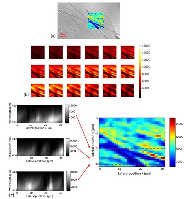
We are currently continuing work on a non-axial-scanning second harmonic imaging technique, in which the chromatic aberration of a Fresnel lens is exploited to focus different wavelengths of a fundamental beam into different axial positions to effectively realize axial scanning. Since the second harmonic signals at different axial positions are generated by different fundamental wavelengths and hence accordingly have different center wavelengths, they can be resolved and detected in parallel by using a spectrometer without axial mechanical scanning. So far, we have demonstrated a system capable of achieving about 8 μm effective axial scanning range [3].
Figure right: Microscope image of a LiNbO3 crystal scratched by silicon carbide sandpaper showing an “X” shaped scratch on its surface (b) Chromatic second harmonic imaging results; the crystal was scanned in two lateral dimensions (30 μm × 30 μm). A series of second harmonic images at wavelengths from 404 nm to 413 nm with an interval of 0.52 nm were obtained and shown here. (c) Side cross-sectional imaging results; three images correspond to y = 20 μm, 26 μm, and 30 μm respectively. Adopted from Yang et al. (2010) [3].
References
[1] K. Shi, P. Li, S. Yin, and Z. Liu, "Chromatic confocal microscopy using supercontinuum light," Optics Express 12, 2096-2101(2004).
[2] Q. Xu, K. Shi, S. Yin, and Z. Liu, “Chromatic two-photon excitation fluorescence imaging,” Journal of Microscopy 235, 79-83 (2009)
[3] Chuan Yang, Kebin Shi, Haifeng Li, Qian Xu, Venkatraman Gopalan, and Zhiwen Liu, “Chromatic second harmonic imaging”, Optics Express 18, 23837-23843 (2010).
[4] Wei-Hung Su, Kebin Shi, Zhiwen Liu, Bo Wang, Karl Reichard, Shizhuo Yin, "A large-depth-of-field projected fringe profilometry using supercontinuum light illumination," Optics Express 13, 1025-1032 (2005).
Support
This work has been supported by the National Science Foundation.
The Pennsylvania State University Department of Electrical Engineering